Diabetic Retinopathy
Author: Dr. Guneet J Mann, MD
Diabetes mellitus is a group of metabolic disorders characterized by a high blood sugar level over a prolonged period of time. As of 2019, an estimated 463 million people had diabetes worldwide (8.8% of the adult population), with type 2 diabetes making up about 90% of the cases.
Diabetes is of three types, Type 1 diabetes , which is caused by inadequate insulin production by Beta cells of Pancreas; Type 2 diabetes, which is caused by an inadequate response of the body to the insulin produced; Gestational diabetes, that develops in pregnant women, without a previous history of diabetes. Gestational diabetes usually resolves after the birth of the baby.
Diabetes affects almost all the major organs of the body including the heart, blood vessels, nerves, eyes and kidneys. Its effect on the eyes is called Diabetic Retinopathy, which involves damage to the retina (inner layer of the eye ball that is responsible for vision). Diabetic Retinopathy is a leading cause of blindness in developed countries. It affects up to 80 percent of those who have had diabetes for 20 years or more. The longer a person has had diabetes, the higher the chances of developing diabetic retinopathy. People with a poor glycemic (blood sugar control) control develop retinopathy earlier.
Over time, raised sugar levels in the blood can lead to the blockage of the tiny blood vessels that nourish the retina, the capillaries and arterioles, cutting off its blood supply. New blood vessels grow in response to various growth factors released by the dying retina, in an attempt to supply nutrition to the retina. But these new blood vessels are abnormal and leak easily.

Types of diabetic retinopathy:
Nonproliferative Diabetic Retinopathy (NPDR)
This is the early and the more common form of diabetic retinopathy. The walls of the retinal blood vessels weaken, resulting in tiny bulges (microaneurysms) protruding from the walls of the smaller blood vessels.
Sometimes these leak fluid and blood into the retina. Larger retinal vessels can begin to dilate and become irregular in diameter, as well. In the natural course, NPDR progresses from mild to severe, as more blood vessels become blocked.
With progression, there may also be swelling of retinal nerve fibers. Sometimes there is fluid accumulation in the central part of the retina (macula) which results in a drop in vision. This requires treatment in the form of Laser or injection of a medicine in the eye.
Proliferative Diabetic Retinopathy (PDR)
This is the more severe, advanced stage of diabetic retinopathy. The damaged blood vessels close off, resulting in the growth of new, abnormal blood vessels in the retina-called neovascularization. These vessels can leak (bleed) into the vitreous (clear, jelly-like substance that fills the center of the eye). A large bleed can cause sudden deterioration of vision.
Stimulated by the growth of new blood vessels, scar tissue eventually forms. When the scar tissue contracts, it can detach the retina. This will cause loss of vision in the areas corresponding to the detached retina. If the new blood vessels and the scar tissue interfere with the normal outflow of fluid (aqueous) of the eye, pressure may increase in the eyeball. This can damage the nerve that carries images from the eye to the brain (optic nerve), resulting in glaucoma. If this happens it is called Neovascular Glaucoma.
Risk factors
Anyone who has diabetes can develop diabetic retinopathy. The risk of developing diabetic retinopathy can increase as a result of:
- Duration of diabetes — the longer a patient has had diabetes, the greater the risk of developing diabetic retinopathy
- Poorly controlled blood sugar levels
- High blood pressure
- High cholesterol levels
- Pregnancy
- Tobacco use
- Being African-American, Hispanic or Native American is a risk factor for development of diabetic retinopathy.
Symptoms
Diabetic retinopathy usually affects both eyes. There may be no symptoms in the early stages of diabetic retinopathy. As the condition advances, diabetic retinopathy symptoms may include:
- Spots or dark strings floating in the field of vision (floaters)
- Blurred vision
- Fluctuating vision
- Impaired color vision
- Dark or empty areas in the field of vision
- Vision loss
Screening for Diabetic Retinopathy
As per the American Academy of Ophthalmology, following recommendations are made for screening for diabetic retinopathy:
-
People with Type 1 diabetes should have annual examinations for diabetic retinopathy beginning five years after the onset of their disease.
-
Type 2 diabetics should have an examination for diabetic retinopathy at the time of diagnosis, then at least yearly examinations thereafter.
-
Women who develop gestational diabetes do not require an eye examination during pregnancy, and do not appear to be at increased risk for developing diabetic retinopathy during pregnancy. However, diabetics who become pregnant should be examined soon after conception and early in the first trimester of the pregnancy. The recommended follow-up is every 3-12 months for no retinopathy or moderate nonproliferative diabetic retinopathy (NPDR), or every 1-3 months for severe NPDR.
Diagnosis of Diabetic Retinopathy
Diabetic retinopathy is best diagnosed with a comprehensive dilated eye exam. Special tests may need to be ordered under certain circumstances, these are:
-
Fluorescein angiography: Flourescein is a dye that is injected into a vein in the arm. Then pictures are taken of the back of the eye with a fundus camera. As the dye circulates in the blood vessels of the retina, any vessel that is closed , broken down or leaking is picked up in the pictures.
-
Optical coherence tomography: This test provides images that give a cross-sectional view of the retina. It shows the thickness of retina, which helps determine if fluid has leaked into retinal tissue. Follow up images of retinal thickness on OCT can be used to assess effectiveness of the treatment in reducing the fluid collection.
Complications
Since diabetic retinopathy may cause leakage of fluid, lipids or blood, along with the abnormal growth of blood vessels in the retina, its complications can lead to serious vision problems:
- Vitreous hemorrhage. Bleeding from the abnormal new blood vessels into the vitreous jelly may cause floaters (floating dark spots in front of eye) if the bleed is small, or a sudden complete blockade of vision in more severe cases. The blood usually clears from the eye within a few weeks or months.
- Retinal detachment. The scar tissue that grows with abnormal blood vessels can pull the retina away from the back of the eye. This may cause floaters or flashes of light or may even produce severe visual loss.
- Glaucoma. New blood vessels may grow in the front part of the eye and interfere with the normal drainage of fluid from the eye, causing an increased eye pressure. Prolonged increase in eye pressure can damage the optic nerve resulting in neovascular glaucoma.
- Blindness. Eventually, diabetic retinopathy, glaucoma or both can lead to complete and irreversible loss of vision.
Prevention and treatment of Diabetic Retinopathy
Diabetic retinopathy can’t always be prevented. Careful management of diabetes is the best way to prevent vision loss. Regular eye examination, and early intervention for vision problems can help prevent severe vision loss. Since pregnancy may worsen diabetic retinopathy, the eye doctor may recommend additional eye exams throughout the pregnancy.
Prevention:
Risk of getting diabetic retinopathy can be reduced by doing the following:
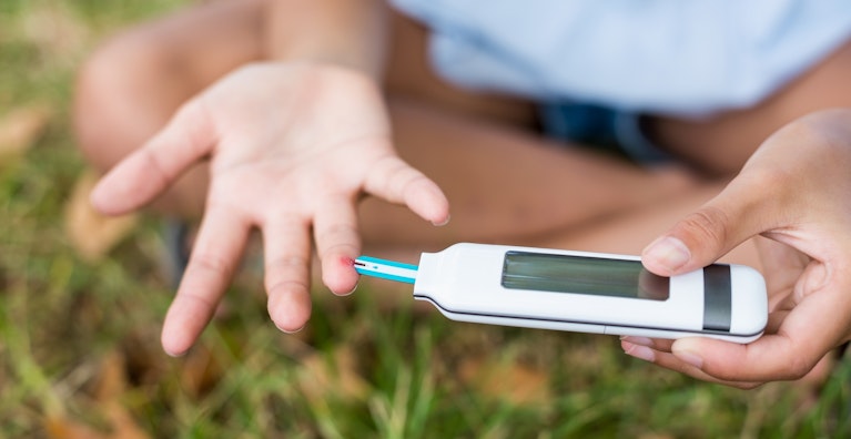
- Adequate management of diabetes: Tight glycemic control can be achieved with dietary modifications, blood sugar lowering medicines and exercises, under supervision of a physician. Assessment of diabetic control should be done with home monitoring of blood sugar levels along with periodic check up of glycosylated hemoglobin (HbA1C) levels
- Keeping blood pressure and cholesterol under control
- Quiting smoking or use of other types of tobacco: Smoking increases your risk of various diabetes complications, including diabetic retinopathy.
- Paying attention to any vision changes. Consult your eye doctor right away if you experience sudden changes in vision or vision becomes blurry, spotty or hazy.
Treatment of Diabetic Retinopathy:
In the early stages of diabetic retinopathy, all that needs to be done is a periodic comprehensive dilated eye examination. The frequency of eye check up is decided based on the severity of the retinopathy. Some people with diabetic retinopathy may need an eye exam as frequently as every 2 to 4 months. In the later stages, some form of treatment needs to be started, more so if there is a change in vision. The treatment modalities are:
-
Intravitreal injections: Small quantities of the medicine are injected into the vitreous cavity of the eye. The drugs commonly used are corticosteroids and Anti VEGF drugs like ranibizumab, bevacizumab etc. Corticosteroids are used for Diabetic Macular Edema (DME) as intravitreal injections, eye drops and implants. The implants could be short acting (Ozurdex-dexamethasone) or long acting (Iluvien-fluocinolone acetonide). Anti-VEGF drugs are used for DME as well as for proliferative diabetic retinopathy, two vision-threatening forms of diabetic retinopathy.
-
Lasers: Lasers are used for causing regression of new blood vessels seen in proliferative retinopathy and also DME. The type of Laser treatment used depends on the indication for which the Laser is used. Panretinal photocoagulation (PRP) is used for proliferative diabetic retinopathy; grid treatment is used for macular edema; and focal treatment seals up small areas of leakage in the retina.
-
Vitrectomy: In vitrectomy the vitreous is cut with a vitreous cutter and most of it is suctioned out. This surgery is done when there is a dense vitreous hemorrhage (bleeding in vitreous) that causes a drop in vision and is unlikely to clear on its own. It is also done in the advanced stages of proliferative diabetic retinopathy when the scar tissue associated with the new blood vessels contracts and detaches the retina. It may be combined with Laser or cryo (freezing) to reattach the retina. Sometimes an air bubble or a special gas is injected into the eye to hold the retina in place. The vitreous fluid may be replaced by a clear fluid, like silicone oil.
The fact is, diabetes doesn’t necessarily lead to vision loss. By taking an active role in the management of diabetes you can go a long way towards preventing complications.
Source:
Share this on Social media
HEALTH DISCLAIMER
This blog provides general information and discussions about health and related subjects. The information and other content provided in this blog, or in any linked materials, are not intended and should not be construed as medical advice, nor is the information a substitute for professional medical expertise or treatment.
The content is for information purpose only and is not a medical advice. Qualified doctors have gathered information from reputable sources; however Credence Medicure Corporation is not responsible for errors or omissions in reporting or explanations. No individual should use the information, resources and tools contained herein to self diagnose or self treat any medical condition.
If you or any other person has a medical concern, you should consult with your health care provider or seek other professional medical treatment. Never disregard professional medical advice or delay in seeking it because of something that have read on this blog or in any linked materials. If you think you may have a medical emergency, call your doctor or emergency services immediately.
The opinions and views expressed on this blog and website have no relation to those of any academic, hospital, health practice or other institution.
Credence Medicure Corporation gives no assurance or warranty regarding the accuracy, timeliness or applicability of the content.
comments powered by Disqus

